We use state-of-the-art imaging techniques to precisely diagnose and effectively treat medical conditions.
The range of services includes X-ray, CT, MRI, and ultrasound – gentle procedures that allow for quick and accurate diagnoses and, in many cases, provide an alternative to invasive methods.
Our partner at the Tamina Health Center is Radiologie Südost, a network of experienced specialists in diagnostic imaging. Their comprehensive services include advanced procedures such as MRI (including neuro-MRI), X-ray, ultrasound, computed tomography, mammography, and osteodensitometry – ensuring accurate and reliable diagnostics.
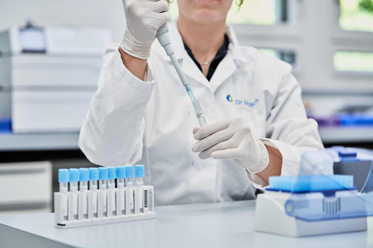
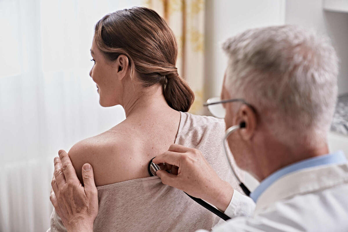
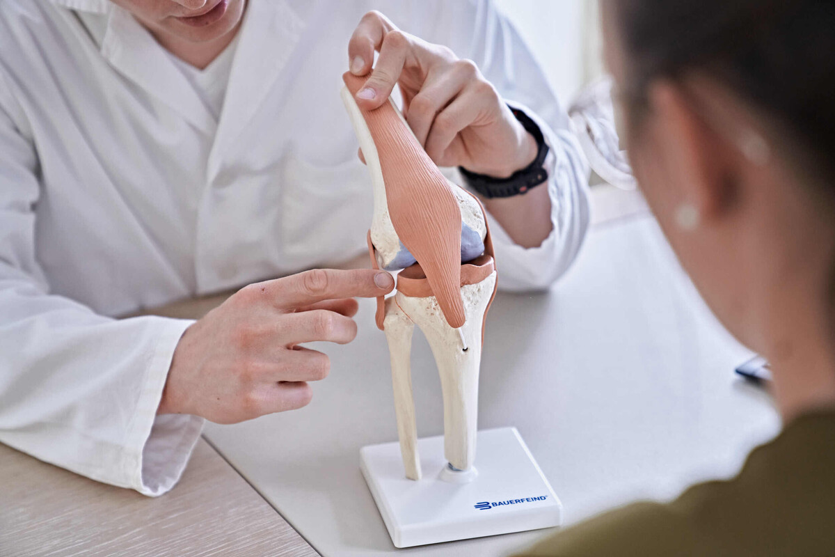
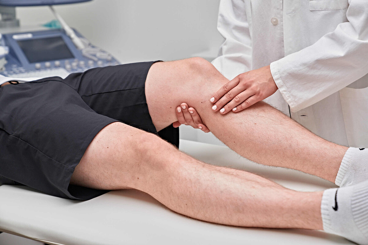
Our Services
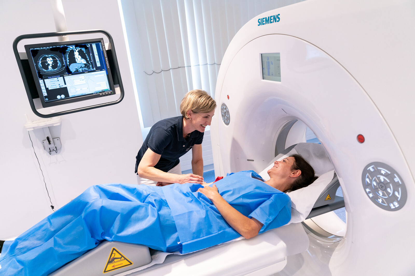
Computed tomography (CT) is a cross-sectional imaging technique and an advanced form of X-ray technology. It is primarily used to examine the head, lungs, abdominal organs, spine, and bones.
How does a CT scan work?
During the procedure, an X-ray tube rotates around the body on a circular gantry, producing a narrow X-ray beam. This beam passes through the targeted body region and is absorbed at different levels by various structures such as fat, muscles, organs, and bones. On the opposite side of the X-ray tube, multiple sensors (detectors) capture the attenuated signal. The result is a highly detailed, multidimensional view inside the human body.
Bone densitometry measures the density and mineral content of bones, allowing for the diagnosis of osteoporosis and the monitoring of treatment success.
How does bone densitometry work?
During the examination, low-intensity X-rays are passed through the body and bones, and the degree of attenuation is measured. This enables precise determination of bone mass and mineral content, which in turn provides information about bone density. Reduced bone density indicates an increased risk of fractures. Measurements are typically taken at the lumbar spine and hip. The radiation exposure is extremely low and considered negligible.
MRI is considered the gold standard for examining the brain, spine, joints, and soft tissues. This modern imaging technique produces detailed cross-sectional images of any body region in multiple planes.
How does MRI work?
Unlike X-ray–based methods, MRI uses a strong magnetic field and radio waves instead of radiation. The signals received are processed by a computer to generate highly detailed images. These images allow for precise differentiation between healthy and pathological changes in the body, particularly in soft tissues such as the brain or internal organs.
Mammography is a specialized X-ray examination of the breast and plays a key role in the early detection of breast cancer. The earlier breast cancer is diagnosed, the better the chances of successful treatment.
How does mammography work?
Mammography is performed using dedicated X-ray equipment. The radiation used is "soft" X-ray, which produces images with higher contrast. This allows even subtle differences in tissue density and composition to be detected. Tiny calcifications (microcalcifications), often an early sign of breast cancer, can also be identified with this method.
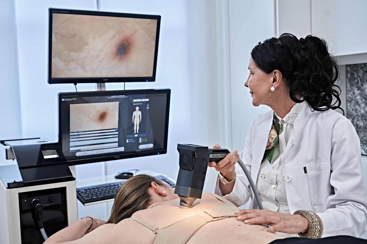
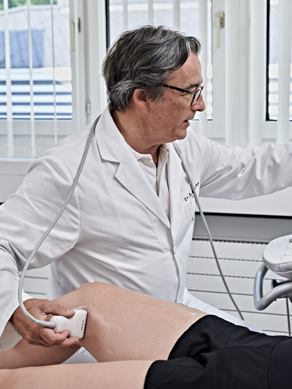
Ultrasound technology is primarily used to examine abdominal and pelvic organs (such as the liver, gallbladder, pancreas, spleen, kidneys, and bladder), soft tissues (including the thyroid and breast), joints, and blood vessels.
How does ultrasound work?
This examination is performed without X-rays. High-frequency sound waves, inaudible to the human ear, are emitted and reflected differently by each organ. The transducer receives the reflected waves, converts them into electrical signals, and transmits them to the ultrasound device, where they are amplified and displayed on a monitor.
With advanced color Doppler (duplex sonography) techniques, the examiner can also obtain detailed information about the speed and direction of blood flow in vessels. This allows vascular conditions such as arterial narrowing (arteriosclerosis) or venous blockages (thrombosis) to be detected.
Conventional X-ray imaging is typically used as an initial assessment of bones, as well as the lungs and heart. Exposing an X-ray image takes only a fraction of a second.
Our team of experts
Contact & Consultation
Schedule an appointment – we’ll be happy to advise you on our services:
+41 81 303 38 61
radiologie.ragaz@hin.ch